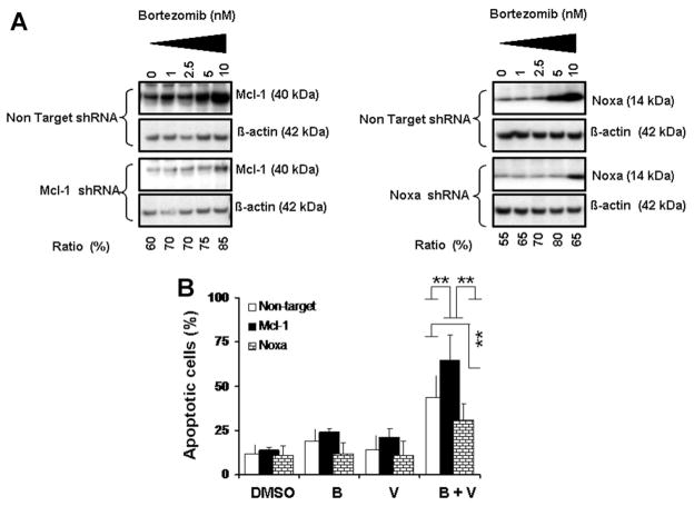
Figure 5.
Knocking down Mcl-1 expression increases glioma cell sensitivity to bortezomib and vorinostat and knocking down Noxa expression protects cells from killing induced by the combination of bortezomib and vorinostat. (A) T98G cells were transfected with non-target or Mcl-1 or Noxa shRNA as described in the Materials and Methods Section. Forty-eight hours posttransfection, cells were treated with the indicated concentrations of bortezomib for 24 h. Lysates were collected and protein was subjected to Western blot analysis using anti-Mcl-1 (left panel) or anti-Noxa (right panel) antibodies. Blots were stripped and reprobed with β-actin. Densitometric analysis of the band intensity was measured, and the numbers below the blot represent percent inhibition of protein in relation to the corresponding control (non-target shRNA). (B) T98G cells were transiently transfected with non-target or Mcl-1 or Noxa shRNA as described in the Materials and Methods Section. After 48 h posttransfection cells were exposed to bortezomib (5 nM) with or without vorinostat (2 μM) for 24 h and cell viability was assessed by annexin V apoptosis assay. Control cells received vehicle (DMSO). Data are representative of triplicate in three independent experiments (**P < 0.005).

