Fig. 2.

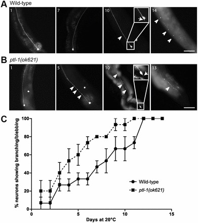
ptl-1(ok621) mutant strain displays an accelerated onset of appearance of abnormal neuronal structures in touch receptor neurons. A representative animal is shown for each genotype (n = 15 total). Neurons were visualised using the Pmec-4::gfp reporter. Worms were imaged every day until death. Representative time points are shown, with arrowheads indicating blebbing and arrows indicating branching phenotypes. For images taken at all time points of a representative worm, see supplementary material Fig. S1. Insets on day 10 show a close-up of the cell body for the respective animal. (A) ALM neuron of a wild-type worm. The worm died on day 15. (B) ALM neuron of a ptl-1(ok621) mutant worm. The worm died on day 14. (C) Percentage of branching/blebbing observed in the complete data set of wild-type and ptl-1(ok621) animals (n = 15). Values are mean±s.e.m. A paired t-test was applied to the means of each strain at each time point and the average of the entire curve compared; P<0.001. Scale bars: 50 µm.
