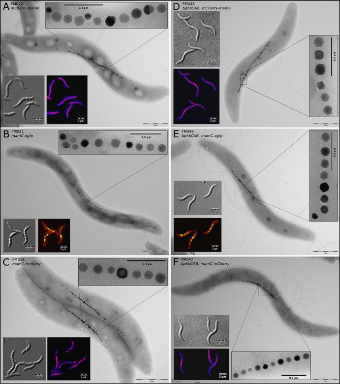
FIG 3.
Differential interference contrast, fluorescence, and TEM images of MSR-1 wt (left column) and phbCAB mutant (right column) cells with markerless chromosomal fusions of mamC and mamK to mCherry (red) and egfp (green). The filamentous fluorescence signals are of even intensity throughout the cell populations (see also Fig. S5 and S6 in the supplemental material). All strains display wt-like magnetosome chains and crystals, indicating that the fusion proteins are functional and that there are no polar effects on downstream genes. (A and D) mCherry-mamK; (B and E) mamC-egfp; (C and F) mamC-mCherry. Membranes in panels B and E were stained with FM4-64 (red), and those in panels A, C, D, and F were stained with Cellbrite Blue cytoplasmic membrane stain.

