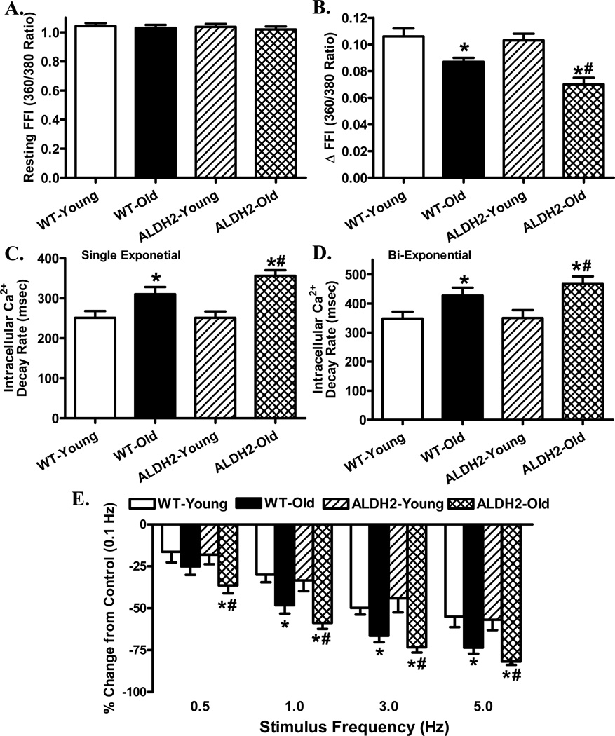
Fig. 1.
Intracellular Ca2+ and frequency response in cardiomyocytes from young or old WT and ALDH2 transgenic mice. A: Resting fura-2 fluorescence intensity (FFI); B: Electrically-stimulated rise in FFI (ΔFFI); C: Single exponential intracellular Ca2+ decay; D: Bi-exponential intracellular Ca2+ decay; and E: Changes in peak shortening of cardiomyocytes (normalized to that obtained at 0.1 Hz from the same cell) at various stimulus frequencies (0.1 – 5.0 Hz). Mean ± SEM, n = 65 cells (panels A–D) and 22 – 28 cells (panel E) from 5 mice per group, * p < 0.05 vs. WT-young group, # p < 0.05 vs. WT-old group.

