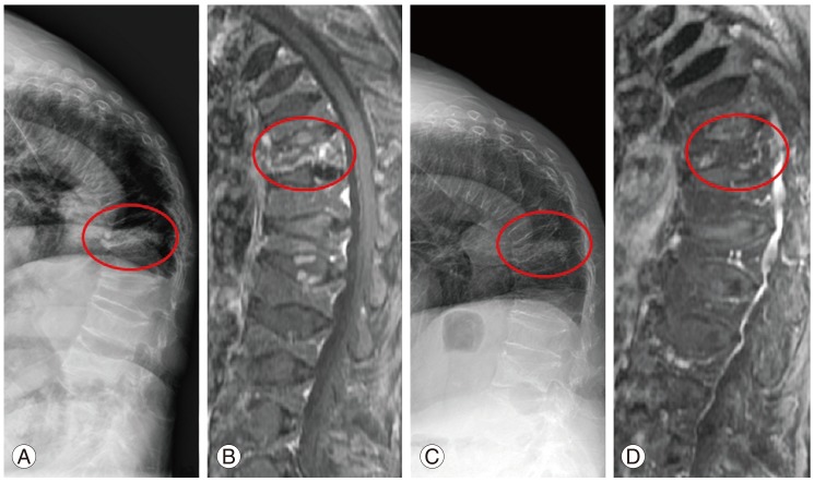
Fig. 1.
(A, B) A 74-year-old female's simple radiograph and magnetic resonance imaging (MRI) demonstrating acute osteoporotic compression fracture in T12, multiple old compression fractures, and resultant kyphosis. (C, D) Simple lateral X-ray and MRI at 3 months after of parathyroid hormone injection show the consolidation of the fracture site.

