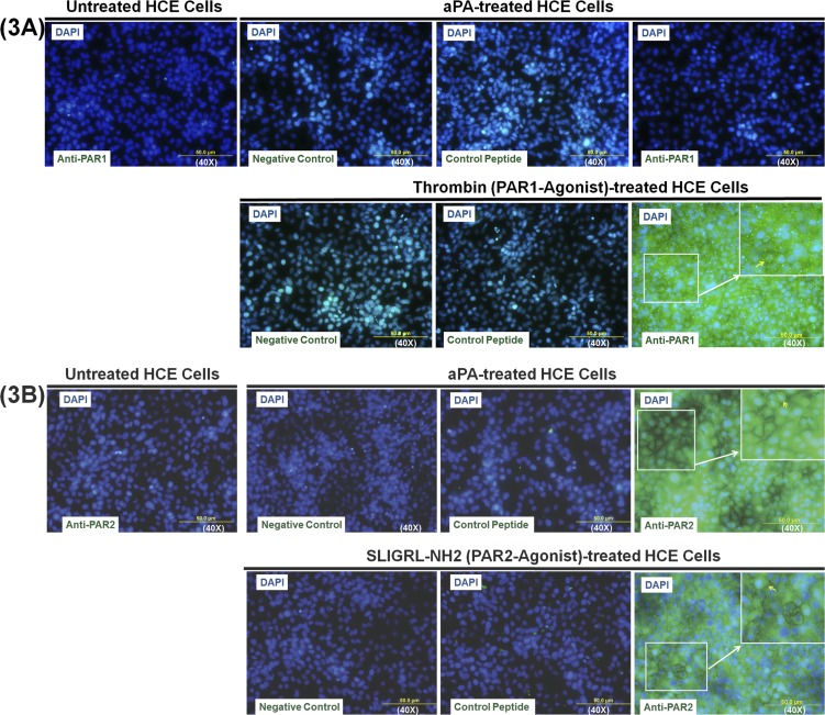
Figure 3.
Acanthamoeba plasminogen activator–induced upregulation of surface PAR2 expression in HCE cells. HCE cells were incubated with or without aPA (100 μg/mL), PAR1 agonist (thrombin, 10 μM), and PAR2 agonist (SLIGRL-NH2, 100 μM) for 24 hours. PAR1 (A) and PAR2 (B) surface protein expression in HCE cells was examined by immunocytochemistry using polyclonal rabbit anti-human PAR1 antibody, anti-PAR2 antibody, and Alexa Fluor 488-conjugated anti-rabbit antibody. Cells without primary antibody incubation were used as a negative control. Human PAR1 (61–76) peptide and rat PAR2 (368–382) peptide were used as absorption control (to demonstrate that PAR1 and PAR2 antibody are binding specifically to the antigen of interest). DAPI counterstaining was used to visualize cell location and morphology. Three slides in each group were viewed using fluorescence microscopy. Images were captured with an Olympus AX70 upright compound microscope.

