Figure 3.
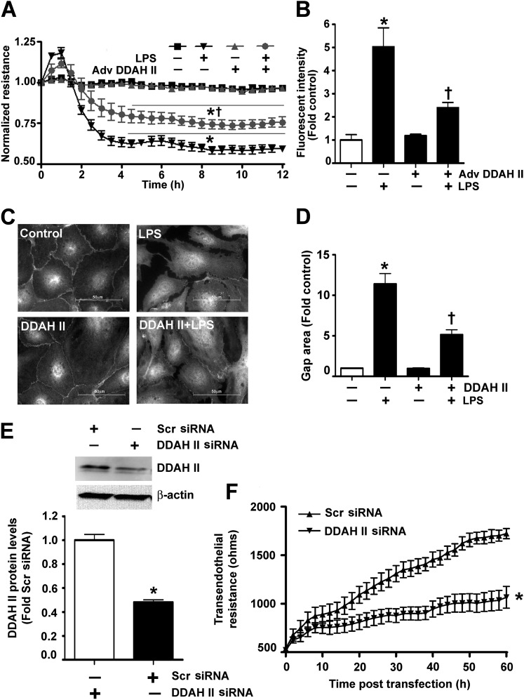
DDAH II overexpression prevents the LPS-induced barrier disruption in HLMVECs. HLMVECs were transduced with Adv GFP (as a control) or Adv DDAH II. After 48 hours, the cells were challenged with LPS (1 EU/ml, 0–12 h). TER was decreased in response to LPS in the control cells, whereas, in the cells overexpressing DDAH II, the decrease in TER was significantly attenuated (A). Similarly, the LPS-mediated (1 EU/ml, 4 h) extravasation of FITC-dextran was significantly attenuated in cells overexpressing DDAH II (B). HLMVECs were also transfected with either pcDNA3 (control) or pcDNA3-DDAH II-His6 plasmid (DDAH II), and exposed to LPS (1 EU/ml, 4 h). Immunofluorescent analysis of vascular endothelial–cadherin (C) indicated that the LPS-mediated increase in gap formation was significantly attenuated in His6-tagged DDAH II–expressing cells (D). When HLMVECs were transfected with either a scrambled small interfering RNA (siRNA) or DDAH II siRNA for 60 hours to reduce DDAH II protein levels (E), basal TER was significantly attenuated (F). Values are mean ± SEM (n = 4–5). *P < 0.05 versus control or scrambled (Scr) siRNA, †P < 0.05 versus LPS.

