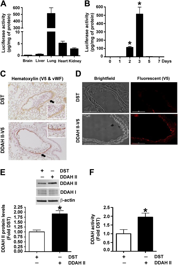
Figure 4.
The overexpression of DDAH II in the mouse lung. The relative amount of luciferase activity was detected in homogenates of brain, liver, lung, heart, and kidney after tail vein injection of pDST-luciferase (40 μg) complexed with polyethyleneimine derivative transfection reagent (Jet-PEI). Luciferase activity was found to be predominantly localized to the lung (A). Maximum luciferase activity in the lung was detected 3 days after injection (B). Mice were injected with either DST-luciferase (DST) or pAD/CMV/V5-DEST-DDAH II (DDAH II-V5) plasmid via tail vein injection, and DDAH II localization was determined. The image in (C) shows hematoxylin staining with V5-tagged DDAH II (V5; red) and vascular endothelial cells labeled with anti-Von Willebrand factor (vWF; brown). The arrows indicate vWF (brown) staining in the control mice, whereas the reddish-brown staining in the V5-tagged DDAH II tissues is a result of a merger of vWF (brown) and V5 (red). (D) Bright-field images (left) of pulmonary vessels and fluorescent images (right) of V5-tagged DDAH II (V5; red). The results from four separate injections show a predominant expression of DDAH II localized to the endothelial layer (C–D). Furthermore, immunoblot analysis demonstrated a significant increase in DDAH II protein levels (E), whereas the increased conversion of N-a-Methyl-(L)-arginine [4,5-3H] to [3H]-L-citrulline demonstrated an increase in DDAH activity, in the lungs receiving DDAH II-V5 plasmid (F). Values are mean ± SEM (n = 4–6 per group). *P < 0.05 versus 0 days (B) and DST (E and F).

