Figure 2.
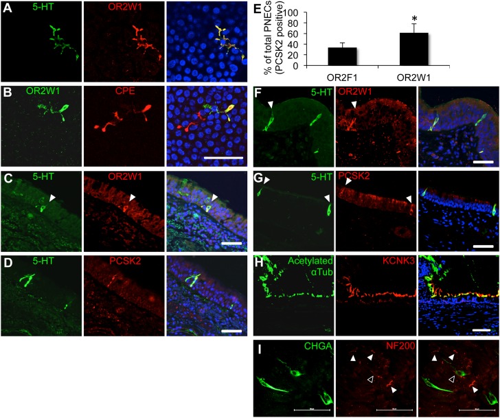
Human pulmonary olfactory cells are solitary pulmonary neuroendocrine cells (PNECs). The OR, OR2W1, is coexpressed with 5-HT (serotonin) (A) and the enzyme, carboxipeptidase E (CPE) (B), in human primary airway epithelial cell preparations. The OR, OR2W1, is coexpressed with the neuroendocrine markers, 5-HT (C) and proprotein convertase subtilisin/kexin type 2 (PCSK2) (D), in human lung tissue sections. (E) OR abundance in preparations from a single donor. Single culture membranes were cut into two pieces and costained for the specified receptor and the PNEC marker, PCSK2. Mean receptor abundance (% cells) is shown for the receptor and PCSK2 out of the total PCSK2-positive cells. Error bars denote SEM. *P < 0.05 (one-tailed t test; n = 3 independent donors). (F and G) PNECs in lung sections from the rhesus macaque. (H) Photomicrographs of the two-pore potassium channel subfamily K member 3 (KCNK3) and the cilia marker, acetylated α-tubulin. (I) Some PNECs might be innervated. Whole-mount immunohistochemistry in the imaged region shows that at least one cell might interact with a neuronal fiber (open arrowhead). Other cells in the imaged area do not seem to interact with neuronal afferents (white arrowheads). PNECs were labeled with anti–chromogranin A (CHGA) antibody (green), and neuronal fibers were labeled with anti–neurofilament H antibody (NF200; red). Scale bars, 50 μm. See Figure E6 for larger images, and Movie E1 for a three-dimensional reconstruction.

