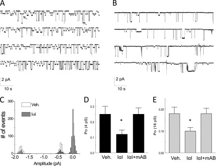
Figure 5.

IαI inhibits ENaC single-channel activity in AT2 cells in mouse lung slices. (A) Cell-attached patches of AT2 cells in lung slices at a holding potential of −100 mV . Note the presence of two Na+ channels with conductances of 4 and 18 pS. Both channels were totally inhibited by the addition of amiloride (10 μM) in the pipette solution (data not shown; see Reference 21). Arrows indicate the simultaneous openings of the 4- and 18-pS channels. (B) A similar record showing decreased channel activity after incubation of the slice with IαI (0.1 mg/ml) for 3 hours. (C) Histogram showing the number of events versus current amplitude (pA) for the records shown in A and B. The corresponding conductances for the three peaks are 4 and 18 pS. Typical records of experiments, which were repeated at least five times. (D, E) Mean values (± 1 SE) (n = 20 for each group) of the open probabilities (Po) of the 4-pS (left panel) and 18-pS (right panel) Na+ channels obtained by patching ATII cells in lung slices incubated with medium (DMEM; Veh), IαI (0.1 mg/ml), or IαI and an equal concentration of an antibody against IαI (mAB IαI; 0.1 mg/ml for ∼ 3 h; *P < 0.0035, as compared with the corresponding control values by one-way ANOVA followed by the Bonferroni modification of the t test for multiple comparisons).
