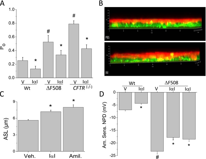
Figure 6.
IαI decreases ENaC activity of lung epithelial cells from wild-type (Wt) and CFTR-deficient mice in vitro and in situ. (A) Lung slices from Wt (Control), ∆F508 (∆F), and CFTR(−/−) mice were incubated with vehicle (V) or IαI (0.1 mg/ml) for 4 hours. Open probability (Po) for the 4- pS channel. Values are means ± SE (n = 20 patches per group; *,#P < 0.001) compared with the corresponding value to the immediate right and the control (Wt) value, respectively; one-way ANOVA followed the Bonferroni modification of the t test adjusted for multiple comparisons. (B, C) Measurements of airway surface liquid volume on murine nasal epithelial cells after addition of IαI (0.1 mg/ml) or vehicle (PBS) for 4 hours. (B) Confocal microscopy figures with the cells labeled with Cell Tracker Green CMFDA and the apical surface fluid with Texas Red (further details are provided in the online supplement). Notice the significant increase of the apical surface liquid on monolayers treated with IαI. Mean values ± 1 SE are shown in C. For comparison, some monolayers were treated with amiloride (10 μM), which completely inhibits ENaC. Number of monolayers used were vehicle (control) = 11; IαI = 11; amiloride = 14 (*P < 0.05 compared with Veh.; ANOVA (P = 0.001) followed by the Bonferroni modification of the t test for multiple comparisons). (D) Measurements of the amiloride-sensitive component of nasal potential difference (NPD) in Wt and ∆F508 mice 4 or 24 hours after intranasal instillation of IαI (0.1 mg/ml; 25 μl in each nostril). Values are means ± 1 SE; number of mice are as follows: Wt (vehicle = 10); Wt IαI = 10; ∆F508 = 20 for each group. Statistical significance is indicated in the graph and was determined as described above.

