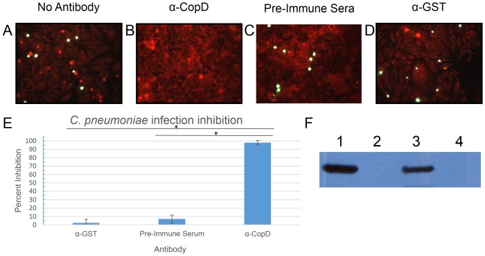
Figure 5. Inhibition of Chlamydia pneumoniae with CopD antibodies.
Panels A–D show inhibition assay results performed with either no antibody (A), CopD antibody (B), pre-immune sera (C), or control antibody (α-GST) (D). Panel E shows the degree of inhibition by of CopD antibodies compared to control antibodies. Chlamydial inclusions are stained green, while HeLa cells are stained red by Evan's blue counterstain. Panel F demonstrates reactivity of anti-CopD with (1) C. pneumonia infected HeLa cell lysate, (2) uninfected HeLa cell lysate, (3) recombinant GST-CopD1–157 produced in E. coli, and (4) recombinant GST produced in E. coli. Experiments were performed in triplicate. Error bars represent 2 standard deviations. Images represent random fields of view. * = P<0.0001.

