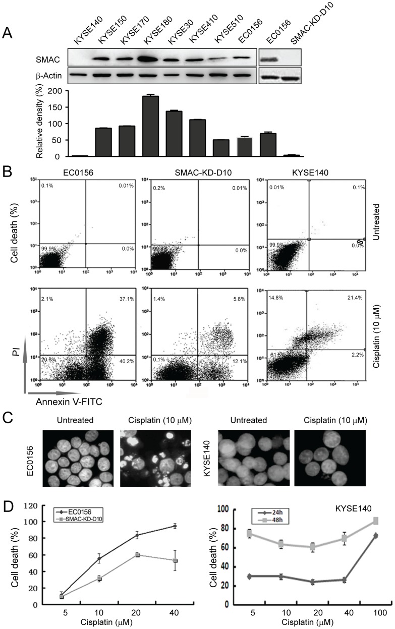
Figure 1. Cisplatin-induced cell death in ESCC cells.
(A) Total cellular protein extracts from each of the nine esophageal cancer cell lines were subjected to western blot analysis for SMAC; SMAC-KD-D10 shows stable knockdown of SMAC. The levels of SMAC protein were quantified, normalized to β-Actin, and are shown in the lower bar graph. (B) Apoptosis was observed in the KYSE140, EC0156, and SMAC-KD-D10 cells after treatment with cisplatin for 24 h, using annexin V/propidium iodide staining and flow cytometry. (C) Nuclei were observed following nuclear staining with DAPI. More apoptotic nuclei appeared in the EC0156 cells after exposure to cisplatin for 24 h, but few apoptotic nuclei were observed in the treated KYSE140 cells. (D) Dose-dependence curve for the KYSE140, EC0156, and SMAC-KD-D10 cells after cisplatin treatment for 24 h and 48 h.

