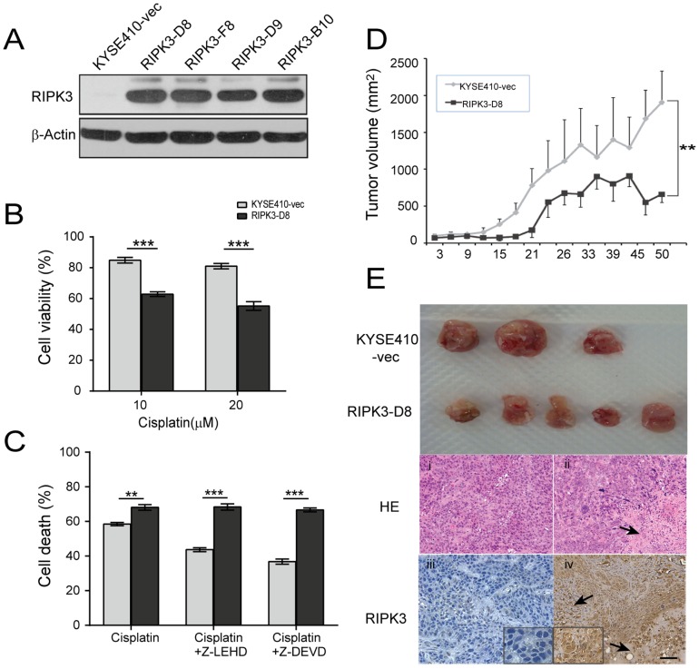
Figure 6. RIPK3 overexpression inhibits tumor growth.
(A) RIPK3 overexpression was analyzed with western blot analysis in the stable KYSE410 clones. (B) RIPK3 overexpression clones and the parental cells were treated with 10 µM or 20 µM cisplatin for 24 h. Cell viability was assessed by flow cytometry. The data represent the mean percentage ± SD of viable cells compared to parental cells. (C) KYSE410-vec and RIPK3-D8 clone cells were treated with 10 µM cisplatin combined with a caspase inhibitor, z-LEHD (5 µM) or z-DEVD (5 µM), for 24 h. Cell viability was determined using an MTT assay. (D) Tumor volume was measured every 2 to 3 days after treatment. RIPK3-D8 clone cells decreased the growth of the xenograft tumors compared to the control group (P<0.001). (E) Reduced tumor growth in the RIPK3-overexpressing cells compared to the parental KYSE410 cells. The parental and RIPK3-D8 cells were suspended in 0.1 ml of PBS and injected into nude mice to establish xenograft tumors. The bottom panels show H&E staining and the RIPK3 expression determined by immunostaining for xenograft tissues. i, H&E staining for control xenograft tissues; ii, H&E staining for RIPK3-D8 xenograft tissue; iii, immunostaining for control xenograft tissues (×100); iv, immunostaining for RIPK3 overexpression in the xenograft tissues (×100). RIPK3 localized mainly in the cytoplasm of the esophageal cancer epithelial cells.

