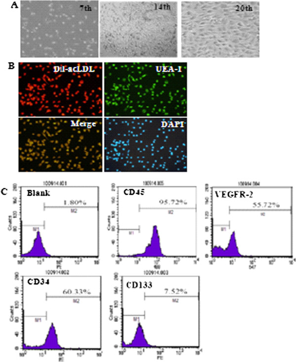
Figure 1.

Characterization of isolated EPCs. A. Representative images of cultured EPCs on 7th day, 14th day, and 20th day (magnification 200×). B. Double-color fluorescent imaging indicated Dil-acetylated LDL incorporation and FITC lectin binding by EPCs. The nuclei was stained by DAPI (magnification 200×). C. FACS showed the expression of stem cell marker CD45, VEGFR-2, CD34, and CD133 in EPCs at 14th day.
