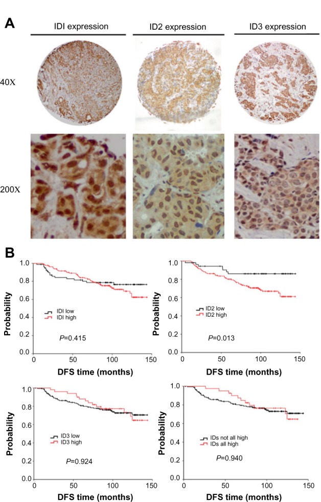
Figure 1.

The expression profiles of ID proteins were determined in breast tumors via immunohistochemistry. (A) Representative photomicrographs of positive immunohistochemical staining for ID1, ID2, and ID3 proteins in breast cancer tissues are illustrated (magnification, 40× and 200×). (B) Kaplan–Meier estimates illustrating DFS, by expression level, for ID1, ID2, ID3 and breast cancer–positive for all ID proteins (including 1, 2, and 3).
Abbreviations: DFS, disease-free survival; ID, inhibitors of DNA binding.
