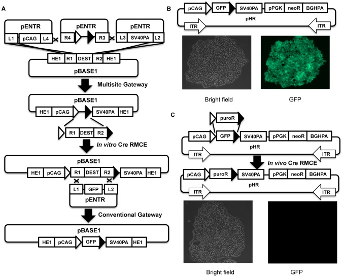
Figure 4. In vitro Cre RMCE cloning and its application in HVAS.
4A, schematic illustration of regeneration of a destination vector with in vitro Cre RMCE cloning. The abbreviations of depicted elements were annotated in Table 1. 4B, fluorescence microscopy for HCT116 cells stably transfected with a 2-module construct built with HVAS and in vitro Cre RMCE cloning as illustrated. Open triangle, canonical loxP; solid triangle, loxN variant. 4C, in vivo RMCE, the cells shown in 4B were transfected with floxed puroR cDNA along with pRN-Cre, selected with both puromycin (1 µg/ml) and G418 (1 mg/ml). Fluorescence and bright field imaging of representative colonies were shown.

