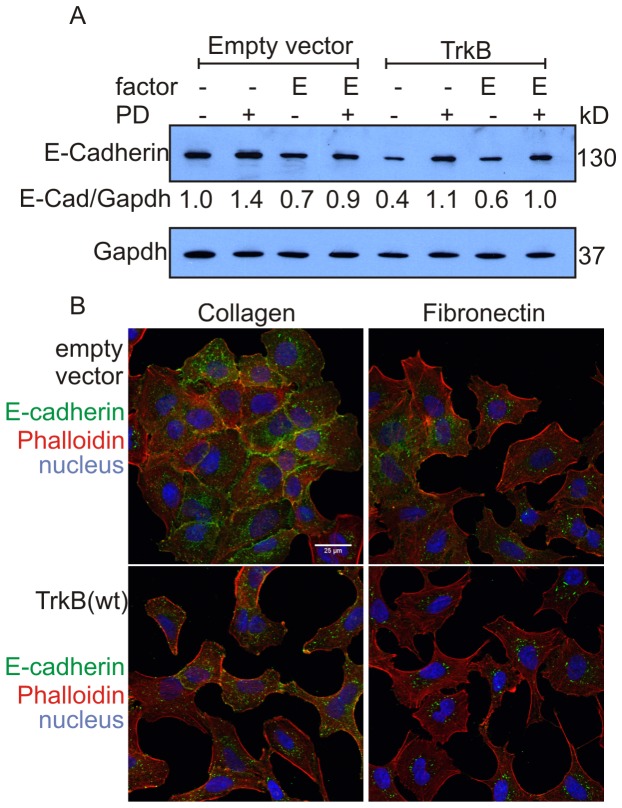
Figure 6. TrkB expression suppresses cell-surface expression of E-Cadherin.
A. Immunoblot analysis of cell lysates from A549 cell clones stably expressing TrkB or harbouring empty vector, cultured on plastic in the absence or in the presence of EGF (E), and with or without the MEK inhibitor PD0325901 (PD), respectively. Expression levels of E-cadherin were quantified by densitometric analysis and normalized by comparison to the Gapdh loading control, shown for each lane as values. B. Immunostaining of E-cadherin and F-actin. Cells were grown on collagen I or fibronectin coated glass slides, serum deprived for 24 hours, fixed and stained. Nuclei were counterstained with DAPI. Results are representative of more than three experiments.

