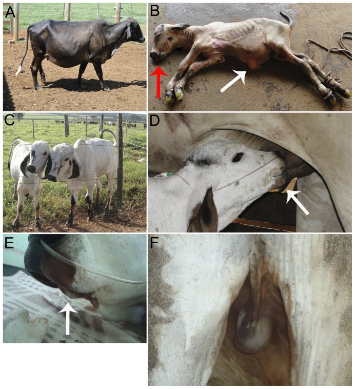
Figure 5. Analysis of health status of cloned calves.
(A) Recipient female from the VPA-treated group showing signals of severe hydroallantois on day 270 of gestation. (B) Stillborn calf from VPA-treated group with enlarged umbilical cord and ascites (white arrow) and brachygnatism (red arrow). (C) Viable calves from the control group. (D) Calf from control group nursing, picture highlighting the correct morphology of the mandible (white arrow). (E) Mandible of cloned calf from control group with moderate brachygnatism (white arrow). (F) Picture highlighting the inguinal region of the calf from the control group evidencing monorchidism.

