
Figure 10.
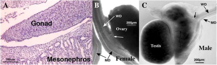
(A) Histological section of an undifferentiated gonad from a 31-day-old fetus sexed by PCR as male. Note absence of seminiferous cords/tubules. (B & C) Wholemount images of spotted hyena genital tracts. (B) Wholemount of a 48-day-old female specimen. Note presence of Mullerian (MD) and Wolffian (WD) ducts and mesonephric tubules (small white arrow). (C) Wholemount of a 50-day-old male specimen. Note presence of Mullerian (MD) and Wolffian (WD) ducts and mesonephric tubules (small black arrow). Both of these specimens exhibit the ambisexual stage based upon differentiation status of the gonads, Wolffian and Mullerian ducts. Subtle texture at the periphery of the testis is indicative of seminiferous tubules, which was confirmed in tissue sections (not illustrated). Adapted from (Cunha et al., 2005) with permission.