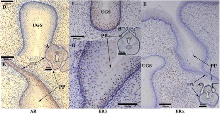
Figure 17.

Androgen receptor (AR) (A, D, E), estrogen receptor beta (ERβ) (B, F, G) and estrogen receptor alpha (ERα) immunohistochemistry (C, E) of a 62-day hyena clitoris. AR was detected in the preputial lamina (PPL), preputial skin and mucosa (PP), and mesenchyme surrounding the UGS (D), but was undetectable in the epithelium of the UGS (F). ERβ was detected in the epithelium of the UGS (F) and preputial skin and mucosa (PP). ERα was detected in the mesenchyme surrounding the UGS (E), but was undetectable in the epithelia of the UGS, preputial lamina, and preputial skin and mucosa (PP).
