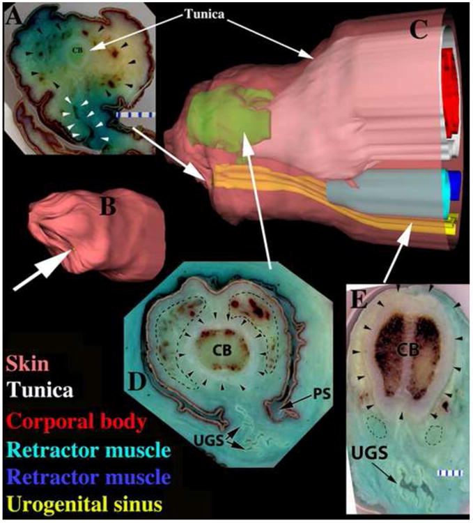
Figure 5.

Three-dimensional reconstructions (3DR) of the clitoris (B-C) as well as sections (A, D, E) of an untreated adult female hyena. (A) Distal section, note corporal body (CB), glanular erectile body (black arrowheads), and UGS (white arrowheads) opening ventrally. Large white arrow pointing to 3DR (B) indicates the position of section (A) in the clitoris. (B) 3DR, ¾ view of the distal end of the clitoris. White arrow indicates urethral orifice on the ventral surface of the clitoris. (C) 3DR, side view with skin semi-transparent to reveal internal structures. Note color-coding of labels and that the retractor muscles are displayed in two different blue hues. (D) Section through the glanular erectile body (dotted lines) as indicated by the large white arrow pointing to a position in 3DR (C). Black arrowheads demarcate the tunica, and dotted lines denote the GEB. Note pleated UGS in ventral position. (E) Proximal section showing corporal body (CB), pleated UGS, tunica (arrowheads) surrounding corporal body only, and retractor muscles (dotted lines) dorsal to the UGS.
