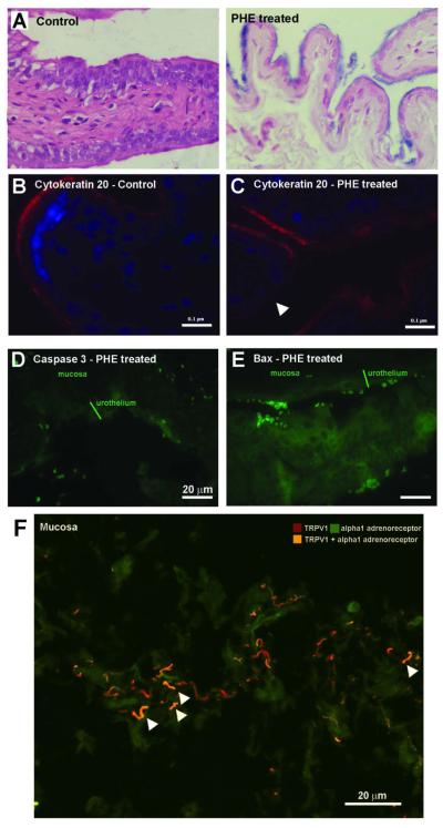
Fig. 3.
(A) Micrograph of a typical control bladder and of a typical area of urothelial thinning following administration of 2.5 mg PHE/Kg body weight, subcutaneously, daily for 14 days. (B) Micrograph showing intense cytokeratin 20 immunolabelling in the umbrella cells of the urothelium of control animals. (C) Micrograph showing areas of decrease or absent cytokeratin 20 immunolabelling (white arrows head) following administration of 2.5 mg PHE/Kg body weight, indicating a loss of umbrella cells. (D) Micrograph showing immunolabelling of the active form of caspase 3 in the urothelium after administration of 2.5 mg PHE/Kg body weight. (E) Micrograph showing bax immunolabelling in the urothelium after administration of 2.5 mg PHE/Kg body weight. (F) Micrograph showing co-localization of alpha 1 adrenoreceptor and TRPV1 imunnolabelling in suburothelial nerve fibres of control animals.

