Fig. 4.
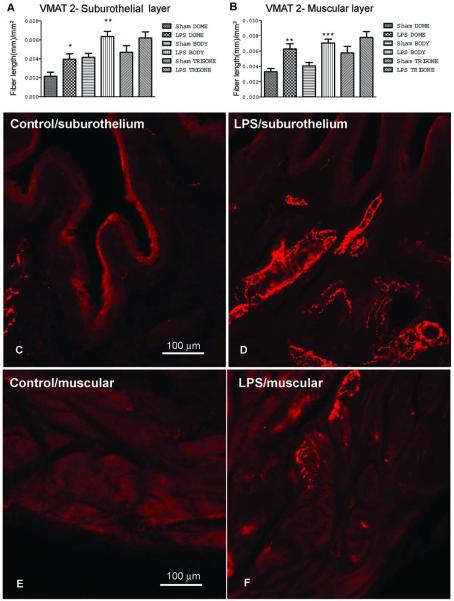
Cystitis induces sympathetic fiber sprouting. Bar graph showing the mean number of VMAT2-immunopositive fiber length/mm2, in (A) suburothelium of the dome, the body and trigone and in the (B) muscular layers of the dome, body and trigone, of both vehicle and 2 mg/kg of LPS treated animals. (*P<0.05, comparing sham suburothelium dome with LPS suburothelium dome, **P<0.01 comparing sham suburothelium body with LPS suburothelium body, and sham muscular dome with LPS muscular dome, ***P<0.001 comparing sham muscular body with LPS muscular body, Kruskal-Wallis test followed by Dunn’s test). Micrographs representative of sections from the dome immunoreactive for VMAT2 are shown in C-F. (C) Bladder suburothelium of vehicle treated animals. (D) Bladder suburothelium of LPS treated animals. (E) Bladder muscular layer of vehicle treated animals. (F) Bladder suburothelium layer of LPS-treated animals.

