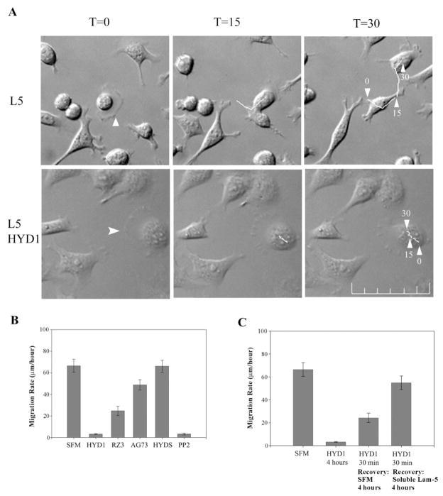
Fig. 1.
HYD1 blocks cell migration on laminin-5. (A) PC3N cells were placed on laminin-5 for 1 h followed by the addition of serum-free media or serum-free media containing HYD1 (75 μg/ml). Videos were started ~30 min post-treatment. Time-lapse images were taken at start of video T = 0, 15 and 30 min. White arrowhead in T = 0 frame marks the cell followed during analysis at T = 30. Scale bar = 100 μm. (B) Cells were added as described in (A) and treated with 75 μg/ml of the peptides HYD1, RZ3, AG73 or HYDS in serum-free media or serum-free media alone (SFM). The Src inhibitor, PP2, was used at a concentration of 10 μM. Videos were started 30 min post-treatment and images were taken every 5 min for 4 h. (C) The effect of HYD1 is reversible. Cells were added as described in (A). Cells were treated with HYD1 for the entire course of the video (4 h). Alternatively, cells were treated for 30 min, the peptide was washed-out, and the cells were exposed to serum-free media alone (SFM) or serum-free media containing soluble laminin-5 and allowed to recover over the 4-h period. Migration rates were quantified as described in Materials and methods. Error bars are standard error from the mean migration rate of 15 cells/experiment.

