
Figure 2.
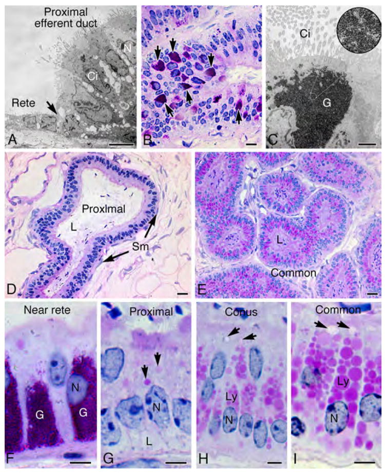
Morphology of efferent ductules in the Golden Syrian hamster. A) Transmission electron microscopy (TEM) of the epithelial junction (arrow) between rete testis and proximal efferent ductule shows cilia projecting into the lumen from the ciliated cell (Ci), while microvilli extend from the surface of the nonciliated cell (N). B) Proximal efferent ductule epithelium near the rete testis contained aggregates of PAS+ granules (arrows). C) TEM of a ciliated cell (Ci) shows aggregates of glycogen (G). At higher magnification in the circular inset photo, the rosettes or glycogen granules are resolved. D) The proximal efferent ductule has a wider lumen (L) than the more distal ducts. E) The common efferent ductule has a narrow lumen (L) and the epithelium contains numerous PAS+ lysosomal granules in the apical cytoplasm. F) Higher magnification of proximal efferent ductal epithelium near the rete testis illustrates an abundance of glycogen (PAS+) granules (G). N, nucleus. G) An area of proximal efferent ductule illustrates epithelial cells with smaller endosomes (arrows) in the apical cytoplasm and lipid-like vacuoles (shown by TEM in Fig. 3 to be large intercellular spaces) in the basal cytoplasm (L). N, nucleus. H) In the conus efferent ductules, nonciliated cells have an abundance of large lysosomal granules (Ly) in the apical cytoplasm and endosomes (arrows). N, nucleus. I) Nonciliated cells in the common efferent duct contain the largest and most numerous lysosomal granules (Ly) and also larger endosomes (arrows). N, nucleus. Bars for A, F-I=5μm; Bar for C=2 μm; Bars for B, D, E=20μm.