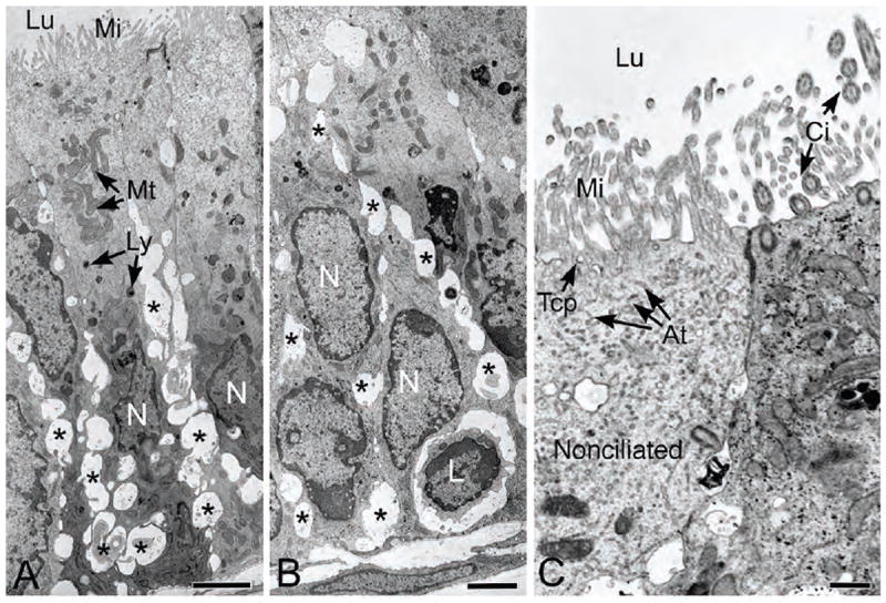
Figure 3.

Transmission electron microscopy of the proximal efferent ductule epithelium. A-B) Numerous basolateral dilations (*) are present in spaces between the epithelial cells. The apical cytoplasm shows numerous mitochondria (Mt) and several small lysosomal-like granules (Ly). The nuclei (N) show considerable variation in shape. One small, lymphocyte-appearing cell (L) is noted near the basement membrane. Lu, lumen; Mi, microvilli. C) Higher magnification of the nonciliated cell (N) reveals initial components of the endocytic apparatus, tubular coated pits (Tcp) and apical tubules (At) beneath a prominent microvillus border (Mi) that lines the lumen (Lu). To the right of the nonciliated cell, cilia (Ci) extend into the lumen from a ciliated cell. Bars for A, B=2.5μm; Bar for C=0.5μm.
