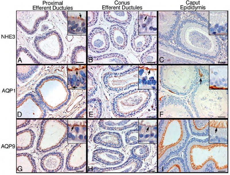
Figure 4.

Immunohistochemistry for sodium/hydrogen exchanger-3 (NHE3), aquaporins-1 (AQP1) and -9 (AQP9) in proximal and conus efferent ductules and caput epididymidis. Arrows point to strongly positive areas of staining. A-C) NHE3 staining was intense on the apical border of nonciliated cells of the proximal efferent ductules, but absent in the ciliated cells. Conus efferent ducts show less intense staining for NHE3, but remain specific for nonciliated cells. Caput epididymidis epithelium was negative for NHE3. D-F) AQP1 staining was intense among the microvilli and cilia of the proximal efferent ducts. Cytoplasmic staining was also observed, as well as along the basolateral membranes. AQP1 staining in the conus region was intense along the microvillus and ciliary borders, but cytoplasmic staining was absent. In the caput epididymidis, AQP1 staining was observed only in capillaries. G-I) AQP9 staining was intense among the microvilli and cilia of the proximal efferent ducts. In the conus ductules, AQP9 showed less intense staining compared to the proximal region. In the caput epididymidis, AQP9 staining was intense along the entire layer of the long microvilli. Bars=50 μm.
