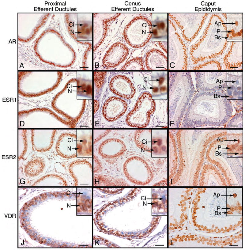
Figure 5.

Immunohistochemistry for androgen receptor (AR), estrogen receptor-α (ESR1), estrogen receptor-β (ESR2) and vitamin D3 receptor (VDR) in proximal and conus efferent ductules and caput epididymidis. A-C) AR was localized as intense nuclear staining in ciliated (Ci) and nonciliated (N) epithelial cells of the efferent ductules and in apical (Ap), principal (P) and basal (Bs) epithelial cells of caput epididymidis. D-F) ESR1 appeared as intense nuclear staining in ciliated (Ci) and nonciliated (N) epithelial cells of the efferent ductules. The cytoplasm also showed immunostaining as well. In caput epididymidis the apical nuclei (Ap) were negative and principal cells (P) showed occasional weak staining, while basal cell nuclei (Bs) had moderate staining. G-I) ESR2 stained ubiquitously all nuclei throughout the efferent ductules and caput epididymidis. Ci, ciliated; N, nonciliated; Ap, apical; P, principal; Bs, basal. J-L) VDR staining was moderate to intense in nuclei of nonciliated (N) epithelial cells of the proximal and conus efferent ductules and in cytoplasm of the proximal ductules. In the caput epididymidis, apical (Ap), principal (P) and basal cells (Bs) were positive for VDR. Bars for A-I=50 μm; Bars for J-L=25 μm.
