
Figure 2.
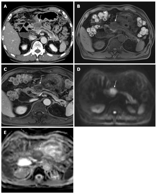
A 73-year-old male with pathologically-proven pancreatic head cancer. A: Approximately 3 cm low attenuating mass (arrow) is noted at the pancreatic head on the CT scan; B: In pre-contrast T1-weighted gradient echo sequence of MR, this mass (arrow) shows lower signal intensity, compared to the normal pancreatic parenchyma; C: After contrast media administration, the pancreatic head cancer (arrow) has poor enhancement; D, E: DWI with 1000 of b-value and ADC map reveal the diffusion restriction of the pancreatic head cancer (arrow). CT: Computed tomography; MR: Magnetic resonance; DWI: Diffusion weighted imaging; ADC: Apparent diffusion coefficient.