
Figure 3.
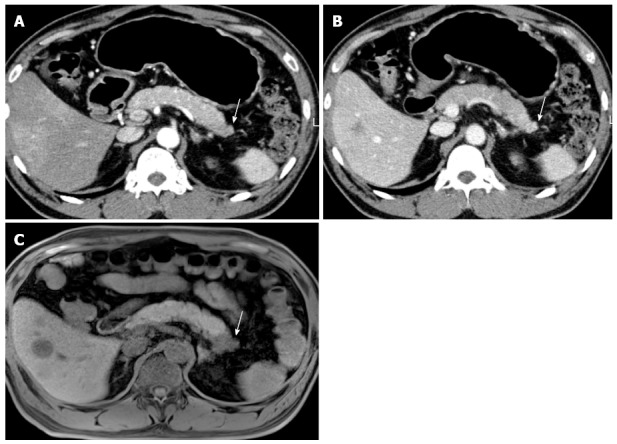
A 64-year-old male with biopsy-proven pancreatic adenocarcinoma with liver metastasis. A, B: On MDCT, the pancreatic tail mass (arrow) shows isoattenuation, causing distal parenchyma atrophy; C: On pre-contrast, T1–weighted, gradient-echo sequence MRI, the pancreatic tail mass (arrow) is clearly depicted, as well as the liver metastasis, owing to the increased soft-tissue contrast of MR compared with that of CT. MRI: Magnetic resonance imaging; MDCT: Multi-detector computed tomography.