Figure 4.
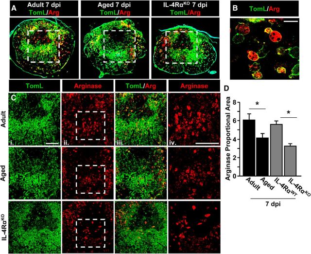
Arginase protein expression was reduced in the injured spinal cord of aged and IL-4RαKO mice. Adult female (3–4 months), aged female (18–22 months), adult male WT (2–4 months), and adult male IL-4RαKO (3–4 months) BALB/c mice were subjected to a T9 laminectomy (Lam) or SCI. Spinal cords were collected 7 dpi. A, Representative images of the injury epicenter in adult, aged, and adult IL-4RαKO mice labeled with TomL (green) and arginase (Arg; red). Dashed white box shows the area represented in C used for quantitative analysis. B, Representative image of TomL/Arg labeling in the epicenter at 63× magnification. Scale bar, 25 μm. C, Representative images of TomL (i), Arg (ii), and TomL/Arg (iii) labeling. Dashed white box shows the area represented in iv at 40× magnification. Scale bar, 200 μm. D, Quantification of Arg staining in adult, aged, adult WT, and adult IL-4RαKO mice (n = 4–6). Error bars represent the mean ± SEM. Means with *p < 0.01 are significantly different from Adult or WT controls.

