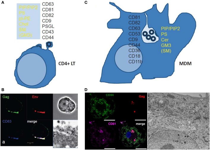
Figure 2.
Scheme and images of HIV assembly at the cellular level. (A) Membrane cell proteins and lipid composition that are found in the virus membrane issued from CD4+ T cell lines or at polarized T cell uropods. From Brügger et al. (2006); Chan et al. (2008); and Lorizate et al. (2013) for the virus lipid membrane composition. From Grigorov et al. (2006, 2009); and Nydegger et al. (2006) for the virus membrane protein composition. From Llewellyn et al. (2013) for the Gag containing T cell uropod microdomains. (B) HIV-1 assembly in chronically infected MOLT cells as shown by immunofluorescence confocal for Gag, Env and the cell CD63 tetraspanin (a) and electron(b) microscopy. (a) HIV-1 infected MOLT cells were fixed in 3% PFA and stained for Gag with an anti-MAp17, anti-gp120, and anti-CD63 for immunofluorescence confocal imaging. (From D. Muriaux lab.) (b) HIV-1 infected MOLT cells were fixed with 2.5% glutaraldéhyde, embedded and thin-sectioned for electron microscopy imaging, as in Grigorov et al. (2009) (From P. Roingeard lab.). (C) The scheme shows the cell membrane proteins and lipids found associated with the viral membrane of HIV-1 issued from monocyte derived macrophages. From Chertova et al. (2006); Chan et al. (2008); Lorizate et al. (2013). (D) HIV assembly in VCC in MDM as shown by immunofluorescence confocal microscopy and electron microscopy. MDM 4 days post-infection with WT HIV-1 NL-AD8, stained by immunofluorescence for CD44 (green), Gag (red), and CD81 (magenta). The nucleus is stained with DAPI. Bar: 20 μm. CD44 and CD81 are present at the plasma membrane but also in intracellular compartments were they co-localize with Gag in infected MDM. (From P.Benaroch lab). For electron microscopy, kindly provided by Mabel Jouve, MDM 15 days post-infection with WT HIV-1 NL-AD8 were fixed, embedded in epon, and ultrathin sections were contrasted with uranyl acetate and lead citrate. Bar: 2 μm.

