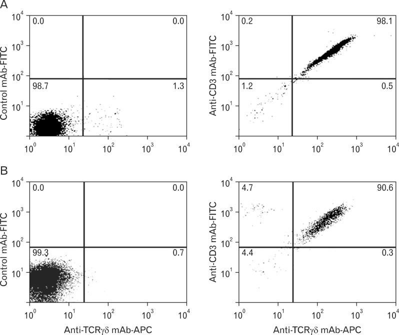
Fig. 1.
Flow cytometric analysis of the purified epidermal and peripheral blood γδ T cells. Purified γδ T cells obtained from human epidermal tissue (A) and from the peripheral blood (B) of healthy volunteers were stained using a fluorescein-conjugated anti-CD3 monoclonal antibody (mAb) and APC-conjugated anti-γδ T cell receptor mAb or the corresponding fluorescently conjugated isotype-matched control Abs. Three independent experiments were performed for epidermal cells and peripheral blood cells respectively, and the representative results are presented. FITC: fluorescein isothiocyanate.

