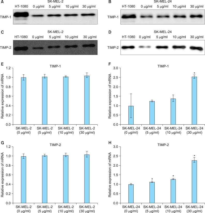
Fig. 5.
Expression of tissue inhibitors of metalloproteinase (TIMP)-1 and -2 protein and mRNA in SK-MEL-2 and SK-MEL-24 cell lines following incubation with imiquimod. Expression of TIMP-1 protein was dose-dependently increased by imiquimod in SK-MEL-2 (A) and SK-MEL-24 cells (B). Expression of TIMP-2 protein was also dose-dependently elevated by imiquimod in SK-MEL-2 (C) and SK-MEL-24 cells (D). Relative mRNA expression of TIMP-1 in SK-MEL-2 (E) and SK-MLE-24 (F) cells and TIMP-2 in SK-MEL-2 (G) and SK-MLE-24 (H) cells, following treatment with a range of concentrations of imiquimod (5, 10, and 30 µg/ml), compared to controls (0 µg/ml). *p<0.05 vs. control.

