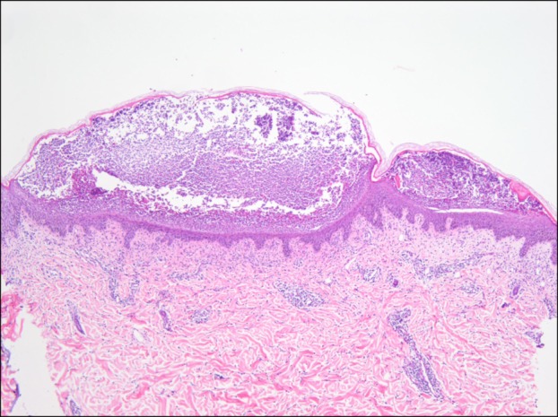
Fig. 2.

Histopathologic findings shows subcorneal pustules in the epidermis, perivascular and interstitial inflammatory cells infiltration in the dermis. The inflammatory cells are comprised of lymphohistiocytes, neutrophils and a few eosinophils (H&E, ×40).
