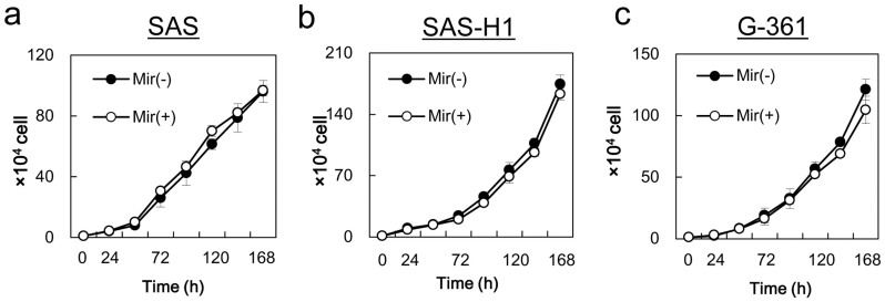
Figure 5. Comparison of cellular proliferation in the control group (without mirtazapine) and the mirtazapine-treated groups.
To determine the effect of mirtazapine on cellular proliferation, SAS (a), SAS-H1 (b), and G-361 (c) cells are seeded in 6-well plates. Cellular proliferation was measured throughout 7 days of treatment. The results are expressed as the means ± the standard error of the mean of values from three assays. Mir (−) = without mirtazapine; Mir (+) = with mirtazapine; h = hours.

