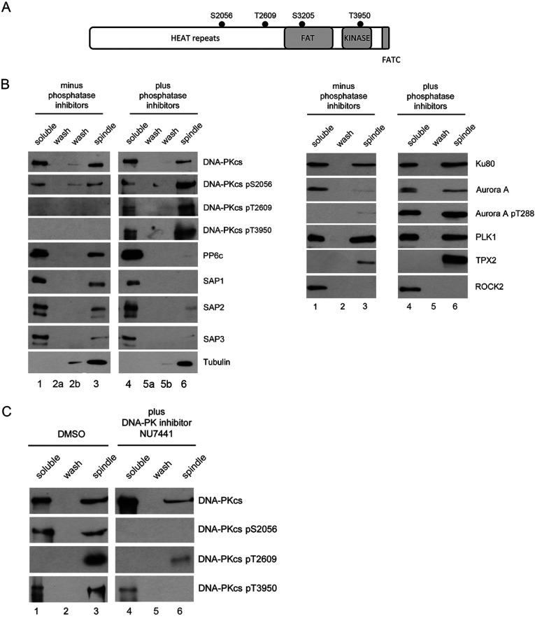
Figure 1. DNA-PKcs binds to and is phosphorylated at enriched mitotic spindles.
(A) Schematic representation of DNA-PKcs showing major domains and positions of Ser2056, Thr2609, Ser3205 and Thr3950 phosphorylation sites. (B) Mitotic spindles and associated proteins were isolated according to [33] either in the absence (left) or presence (right) of protein phosphatase inhibitors (see the Materials and Methods section for details). Lanes 1 and 4 contained soluble proteins. Lanes 2 and 5 contained the low ionic strength wash, and lanes 3 and 6 the mitotic spindle fraction (see the Materials and Methods section for details). In the blots shown in the left-hand panel, an additional low salt wash (lanes 2b and 5b) was included on the gels. Samples were boiled in SDS–PAGE sample buffer, run on SDS–PAGE gels, transferred to nitrocellulose and probed with the described antibodies. (C) Mitotic spindles were prepared as in Figure 1(B) with or without the addition of the DNA-PK inhibitor (NU7441) 1 h prior to mitotic shake off. Samples were run on SDS–PAGE, transferred to nitrocellulose and probed for phosphorylation as indicated.

