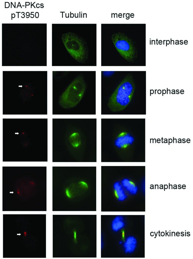
Figure 2. Phosphorylation of DNA-PKcs at Thr3950 in mitotic cells.
U2OS cells were stained with FITC-conjugated α-tubulin (green) at 1:1000 dilution, and phosphospecific antibody to DNA-PKcs phospho-Thr3950 (red) at 1:200 dilution at different phases of the cell cycle. DNA stained with DAPI is shown in blue. An expanded image of a cell in cytokinesis stained with the DNA-PKcs phosphoThr3950 antibody (panel B) is shown in Supplementary Figure S3(A).

