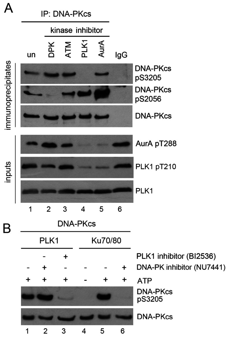
Figure 3. PLK1 phosphorylates DNA-PKcs on Ser3205 in mitosis.
(A) HeLa cells were treated with 40 ng/ml nocodazole for 15 h, then 1 h prior to shake off either mock-treated (un) or treated with 8 μM of the DNA-PK inhibitor (NU7441), 5 μM of the ATM inhibitor (KU55933), 100 nM of the PLK1 inhibitor (BI2536) or 100 nM Aurora-A-Inhibitor-I for 1 h. After shake off, cells were allowed to recover in fresh media, in the absence of nocodazole but in the continued presence of kinase inhibitors, for a further 35 min before harvesting by centrifugation. NETN lysis was carried out and DNA-PKcs was immunoprecipitated from 1 mg of lysate as described in the Materials and Methods section. Immunoprecipitates were blotted with the described antibodies. The lower panel shows 50 μg of the whole cell extract probed for Aurora A Thr288 and PLK Thr210 phosphorylation as well as total PLK1 to show efficacy of PLK1 and Aurora A inhibitors. (B) Purified DNA-PKcs was incubated with His-tagged purified PLK1 alone (lanes 1–3) or in the presence of purified Ku heterodimer (lanes 4–6). Where indicated, reactions contained the DNA-PK inhibitor NU7441 (lanes 2 and 6) or the PLK1 inhibitor BI2536 (lane 3) as indicated. All lanes contained DNA. ATP was present in lanes 1–3 and 5 and 6 as indicated. Reactions were stopped by boiling the samples in SDS sample buffer, and samples were run on an SDS 8%PAGE gel and probed with the described antibodies.

