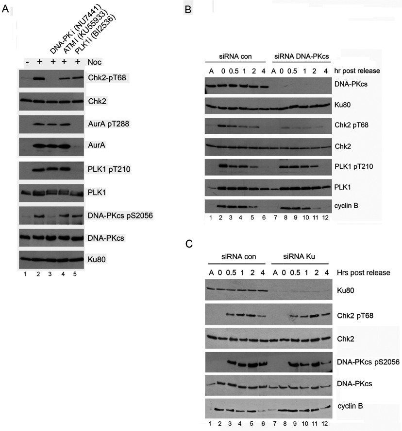
Figure 7. Inhibition of PLK1 does not affect DNA-PK dependent, mitotic phosphorylation of Chk2 on Thr68.
(A) HeLa cells were left untreated or were treated with 40 ng/ml nocodazole for 16 h. One hour prior to mitotic shake off and harvesting, cells were left untreated or were treated with the DNA-PK inhibitor NU7441 (8 μM), the ATM inhibitor KU55933 (5 μM) or the PLK1 inhibitor BI2536 (100 nM). After mitotic shake-off the cells were left to recover for 35 min in the presence of the kinase inhibitors but in the absence of nocodazole. NETN lysates were prepared as described in the Materials and Methods section and 50 μg of extract was resolved on SDS–PAGE gels and probed with the antibodies as described. As in Figure 3(A), inhibition of PLK1 with BI2536 resulted in loss of phosphorylation of both PLK1 and Aurora A. (B) HeLa cells were transfected with siRNA to DNA-PKcs as described in the Materials and Methods section. At 80 h post-transfection, 40 ng/ml nocodazole was added for a further 16 h as indicated or cells were left untreated. Cells were harvested by mitotic shake-off and left to recover in nocodazole-free media for the times indicated. NETN lysates were prepared as described in the Materials and Methods section and 50 μg of extract was resolved on SDS–PAGE gels and probed with the antibodies as described. Quantitation of Figure 7(B) is shown in Supplementary Figure S6. Results are representative of three separate experiments. (C) HeLa cells were transfected with siRNA to siRNA to Ku70/80 and analyzed as described in Panel B. Quantitation of Figure 7(C) is shown in Supplementary Figure S6. Results are representative of three separate experiments.

