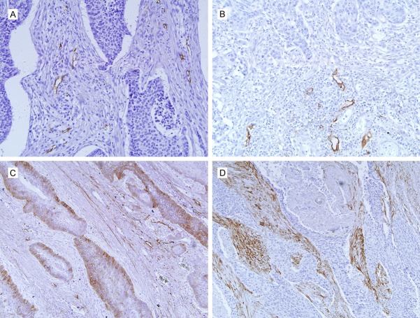
Figure 1.
Podoplanin positive expression in esophageal squamous cell carcinoma. Strong podoplanin staining for lymphatics in intratumoral (A) and peritumoral (B) locations; the blood vessels in peritumoral tissue are negative for D2-40 indicating its specificity for lymphatic vessels. D2-40 positivity is seen in membrane and cytoplasm of cancer cells, and positive cells are mainly located in the periphery of cancer nests (C). (D) Shows stromal podoplanin staining with high amount of podoplanin expressing cancer-associated fibroblasts. Original magnification, ×200.

