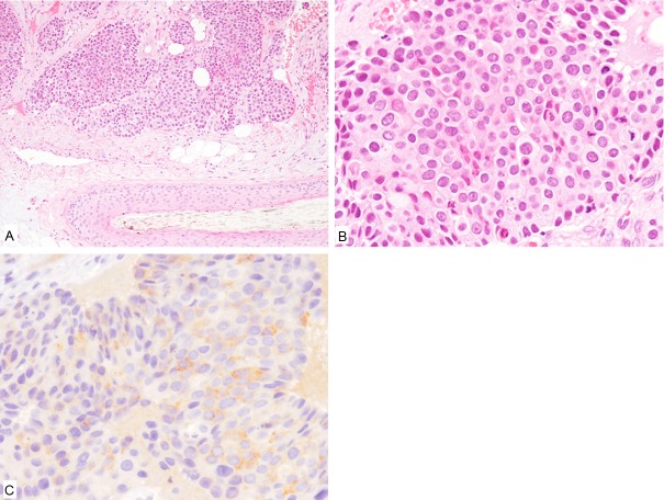
Figure 2.
Histopathological and immunohistochemical features of the scalp nodule. A: Proliferation of variably-sized epithelial nests in the subcutis around the hair follicle. HE, x 100. B: The tumor cells have relatively rich eosinophilic cytoplasm and large round to oval nuclei. HE, x 400. C: Synaptophysin is expressed. x 400.

