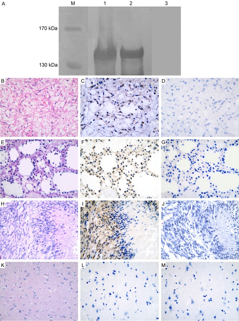
Figure 2.

Specificity analysis of MAGE-D4 antiserum and immunohistochemical staining of MAGE-D4 protein. A: The antiserum can detect the precipitate from induced bacteria containing pMAL-c2/MAGE-D4 plasmid (Lane 1) and purified recombinant MAGE-D4 protein (Lane 2). The molecular weight of immune reactive band was around 140 kDa, which was higher than the predictive value (124 kDa) of MAGE-D4 fusion protein. The pre-immune serum failed to bind with the recombinant MAGE-D4 protein (Lane 3). Lane M, protein marker. C: Cytoplasmic expression of MAGE-D4 in diffuse astrocytoma. F and I: Nucleus and cytoplasm co-expression of MAGE-D4 in diffuse astrocytoma and glioblastoma, respectively. L: MAGE-D4 was negative in normal brain tissue. B, E, H and K: The corresponding tissue sections were stained with hematoxylin-eosin. D, G, J and M: The pre-immune rabbit serum was used as negative control.
