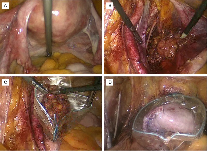
Figure 1.

Laparoscopic features of different steps of surgical staging. A: View of pelvis without macroscopic signs of extra-uterine disease; B: Systematic pelvic lymph nodes removal; C: Abdomen extraction of removed lymph nodes through the ancillary trocar (using the endobag device); D: Abdomen extraction of removed uterus and adnexa through vaginal route (using the endobag device).
