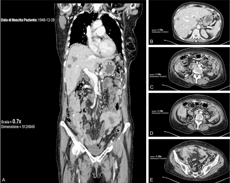
Figure 2.

Computerized Tomography features six month after surgery: nodular multiple signs of lung metastatic lesions, diffuse abdominal carcinomatosis, ascites and multiple metastasis (liver, spleen bowel). A: Intermediate dorsal-ventral coronal imaging. B-E: Progressive cranial-caudal axial imaging of abdomen and pelvis.
