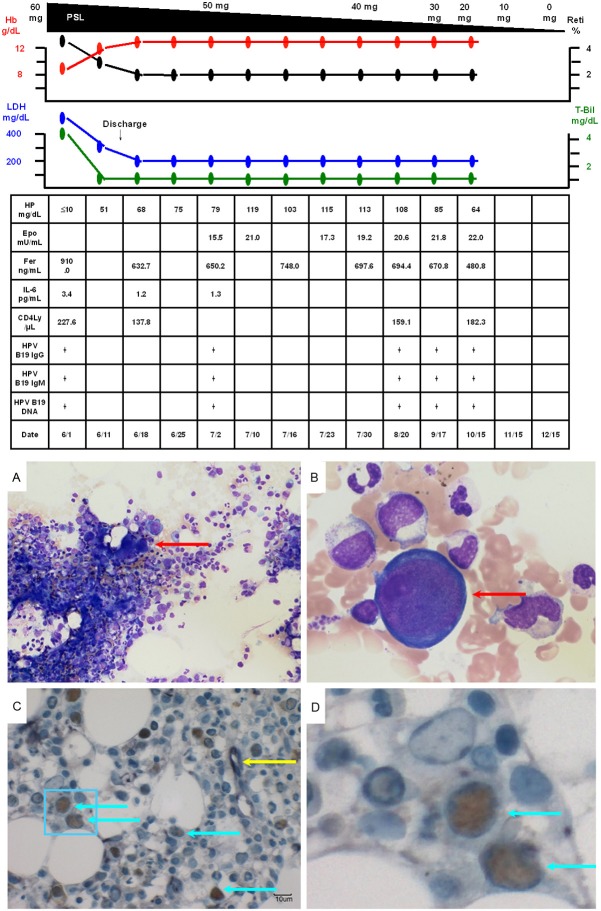
Figure 3.
A. Bone marrow examination (smear; ×40) revealed normal cellularity and a marked decrease in the density of erythroblasts, with an M/E ratio of 117.25. B. Bone marrow examination (smear; ×600) also showed giant proerythroblasts (red arrow), suggesting that the patient also had PRCA. C. Double-immunostaining (enzyme-labeled antibody method; ×400) of bone marrow biopsy specimens showed positivity of the erythroblasts for anti-HPV B19 antibodies (brown staining of nuclei) and anti-erythropoietin receptor antibodies (purple staining of the cytoplasm) (light blue arrows). Some erythroblasts showed positivity for only anti-erythropoietin receptor antibodies (yellow arrow). D. An enlarged image of the area in the light blue frame in C.

