Figure 1.

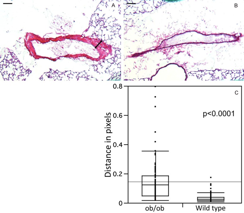
Lung tissues from ob/ob (A) and control (B) lungs were stained using hematoxylin and eosin (H&E). H&E staining demonstrates thickening smooth muscle cell wall and hyperplasia in the ob/ob arteriole (arrow in A) that was not observed in control animals. The p value for the difference in thickness of the arterial wall (distance in pixels) and standard errors were calculated from three different slides by using 10 arteries in each slide. Ten distinct segents of the arterial wall were measured (C). Bar is 100 μm.
