
Figure 5.
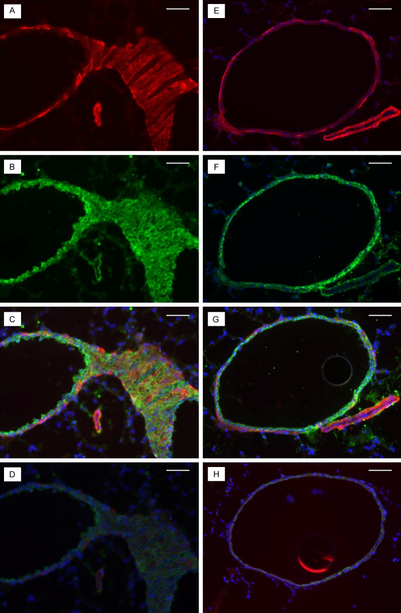
Lung tissues from ob/ob (left) and control (right) lungs were stained for smooth muscle cell actin (SMCs; smooth muscle actin) and HA (HA binding protein). The intensity of SMC actin (red) and HA (green) staining surrounding the ob/ob arteriole (A and B) is greater compared with the control (E and F). C and G are composite pictures of HA and SMC actin for the same sections of ob/ob and control lung tissues, respectively. D and H are negative controls of the same sections stained by the secondary antibody only. The minimal green staining here represents autofluorescence by the elastica, and the blue staining represents the 4,6-diamidino-2-phenylindole (DAPI) staining of the nuclei. Bar indicates 100 μm.