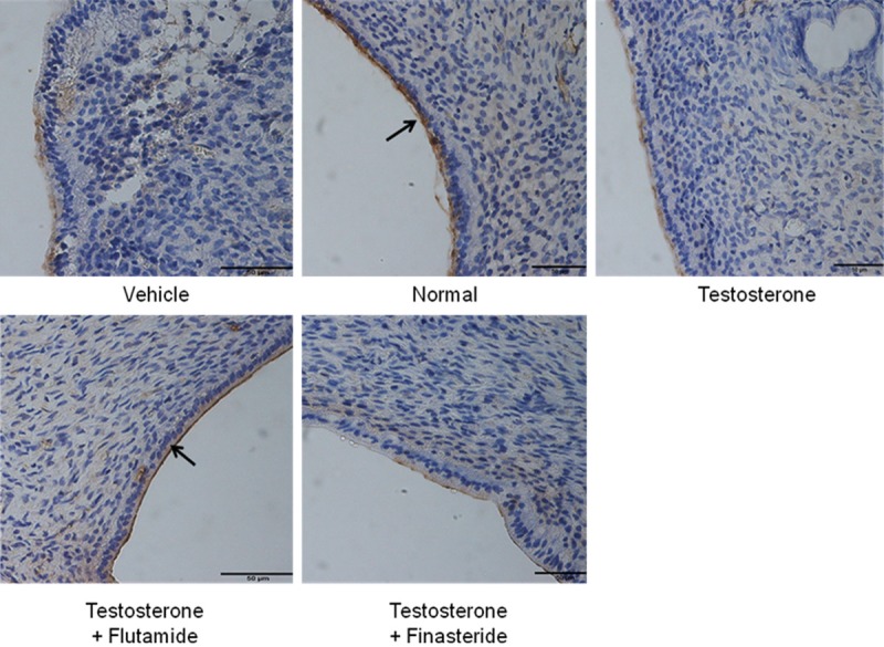
Figure 4.

MECA-79 protein distribution at the apical membrane of the endometrial luminal epithelia in different experimental group. MECA-79 was seen to be distributed at the apical membrane of the luminal epithelia predominantly in rats received normal steroid replacement and concomitant testosterone and flutamide administration during uterine receptivity period. L: lumen, G: gland, scale bar: 50 μm. Arrow pointing towards the apical membrane with highest immunostaining intensity.
